
Electromyography (EMG) is a diagnostic procedure to test the health of the nerve cells that control the muscles in your body. These cells are called motor neurons. They transmit electrical signals that cause muscles to contract and relax. If the motor neurons are not functioning properly, then symptoms like pain, tingling, and numbness might affect parts of the body.
What is Electromyography?
Electromyography is a procedure that measures the electrical activity of the nerves that control muscle function. EMG results can reveal nerve damage and help diagnose disorders related to orthopedic conditions like carpal tunnel, ulnar neuropathy, pinched nerves, and other types of neuropathy. It can also be used in other areas of medicine to diagnose conditions caused by nerve dysfunction.
When is EMG Needed?
A doctor may order electromyography if you have signs or symptoms that may indicate a nerve or muscle disorder. Symptoms that may prompt your doctor to refer you for an EMG include:
- Tingling
- Numbness
- Muscle weakness
- Muscle pain or cramping
- Limb pain
EMG results are often necessary to help diagnose or rule out a number of orthopedic conditions such as:
- Peripheral (outside of the spinal cord) nerve disorders
- Carpal tunnel syndrome
- Ulnar neuropathy
- A pinched nerve in the neck
- Nerve compression
- Muscle disorders
- Muscular dystrophy or polymyositis
- Diseases affecting the connection between the nerve and the muscle
- Myasthenia gravis
- Disorders that affect the nerve root
- Herniated disk in the spine
What Happens During Electromyography
You will need to be referred to an EMG specialist by your doctor to get the test done. Once you have an appointment with the EMG specialist, they will give you specific instructions, but there are some general things you can expect if you have the procedure.
Electromyography is a non-invasive procedure. There is no specific preparation patients need to do before the test. There are no after-effects once the test is complete and no recovery time. So, patients can return to their normal daily activities right after the test is done. Results may take a few days to arrive because they will be sent to your referring doctor for analysis first.
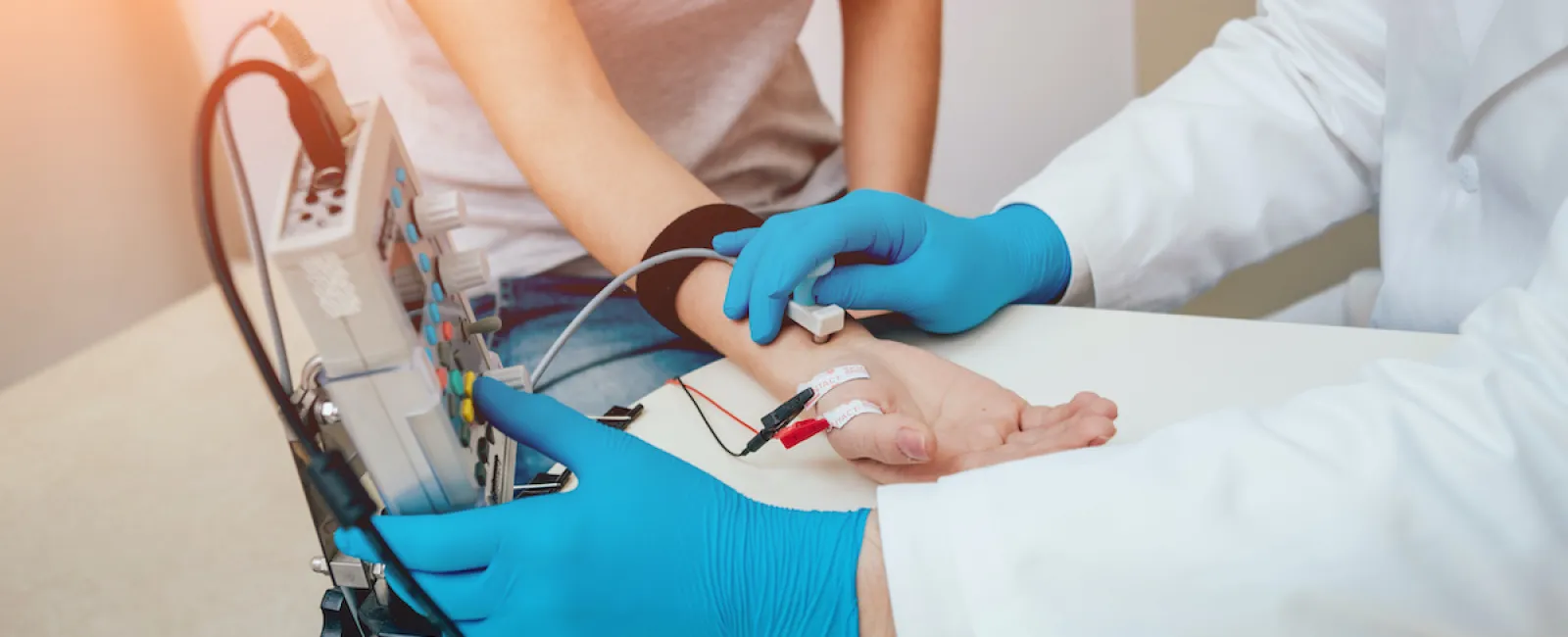
Electromyography tests may include two parts: a nerve conduction study and a needle EMG.
Nerve Conduction Study
A nerve conduction study uses surface electrode stickers applied to the skin to stimulate the nerves. The electrodes then measure the electrical activity of the nerves through the skin. Usually, a technician will perform this part of the test. They will place the electrodes and instruct you on how to position yourself. They will also give you directions on when to move and contract your muscles.
Needle EMG
During a needle EMG, an ultra-thin needle with an electrode on the tip is inserted superficially under the skin in key muscles. The doctor performing the test will monitor the activity of the motor neurons to see if there is any spontaneous electrical activity when the muscle is at rest. A healthy muscle will not have any activity while at rest. They will also look at how much electrical activity happens when you are using the muscle.
Electromyography at South Shore Orthopedics
South Shore Orthopedics is fortunate to offer electromyography with the addition of Dr. Sandra Maguire to our practice. Dr. Maguire is board-certified in Electrodiagnostic Medicine by the American Board of Electrodiagnostic Medicine. If you have symptoms of a nerve disorder and think you’d benefit from diagnostic testing with EMG, call our office at (781) 337-5555 to make an appointment to speak to your doctor about a referral.
